Cone beam computed tomography cbct is a promising modality for quick outpatient imaging with lower radiation dose and less metal artifact when compared to conventional ct mdct scans.
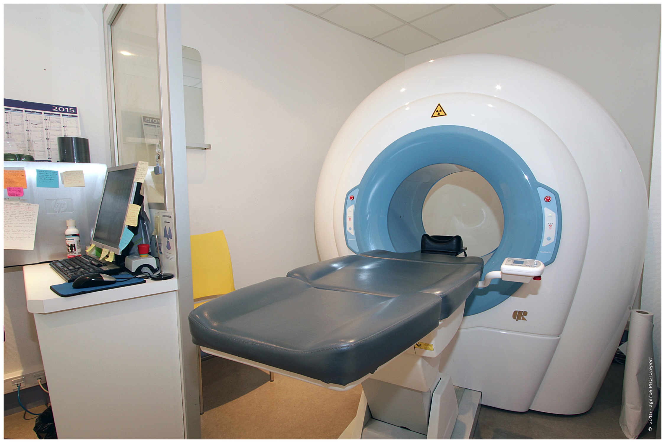
Scanner cone beam sinus.
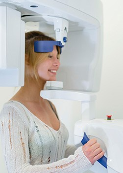
Dental cone beam computed tomography ct is a special type of x ray equipment used when regular dental or facial x rays are not sufficient.
Cone beam ct cbct scanner vs.
This procedure requires little to no special preparation.
Et à l inverse de celui ci il permet de balayer en un seul passage l ensemble du volume à radiographier en étant en outre moins irradiant.
This article will be providing evidence on the diagnostic and treatment planning applications of cbct in sinus imaging mainly.
A cone beam ct scan is not only used to rule out pain of unknown origin or pathology but can also assess their airway for areas of possible airway obstruction.
With continued symptoms patient s endodontist immediately recommended a cone beam ct scan cbct which provides a 3 dimensional view of the site.
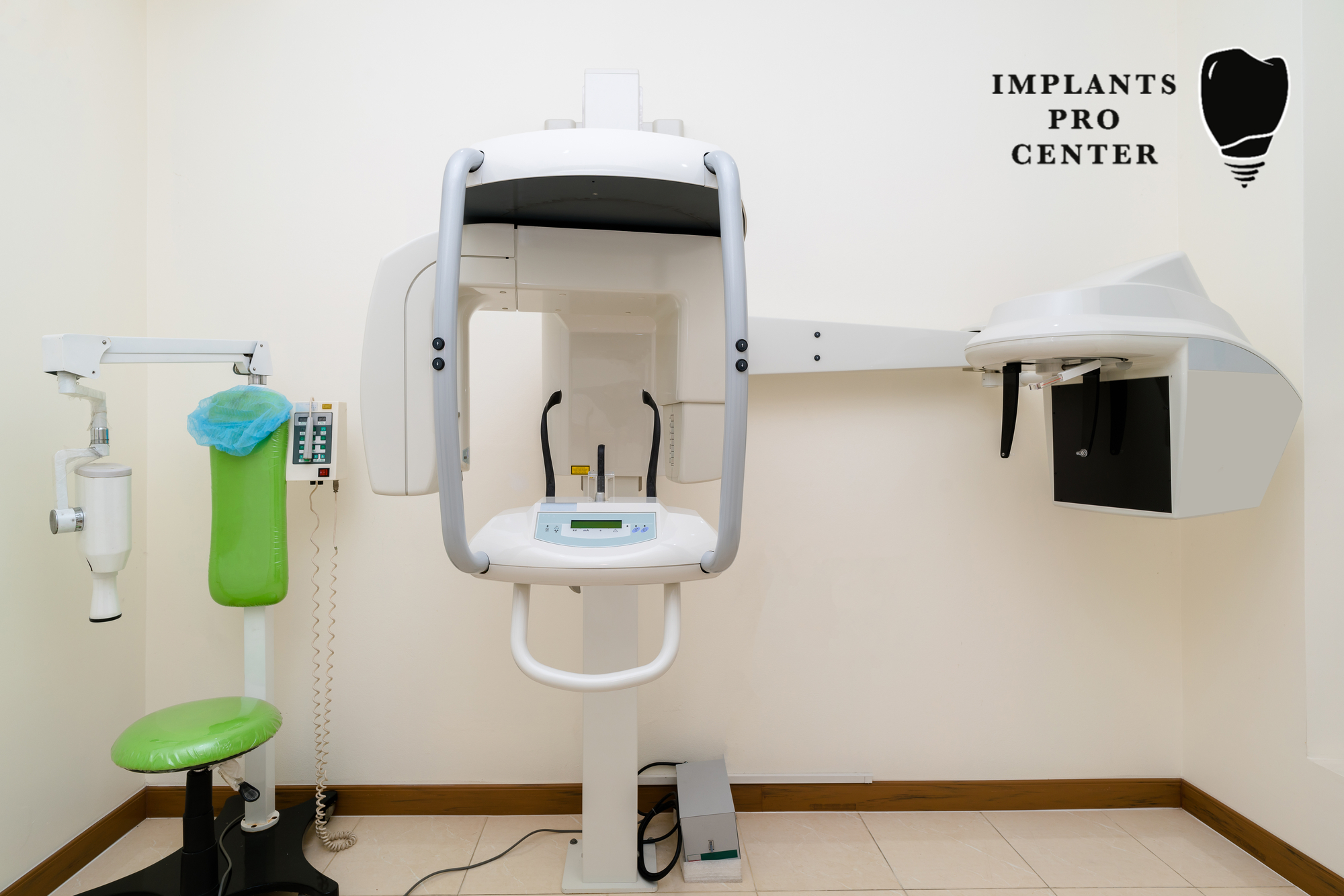
Cbct has become increasingly important in treatment planning and diagnosis in implant dentistry ent orthopedics and interventional radiology among other things.
Your patients will leave their appointment appreciating and knowing they are receiving a thorough examination.
The ct scan used in our office can detect a variety of things including nasal polyps inflammation or infection of the sinuses and fluid filled sinuses.
The scan showed a large area of infection characterized by bone loss measuring 10mm by 9 mm and approaching the maxillary sinus.
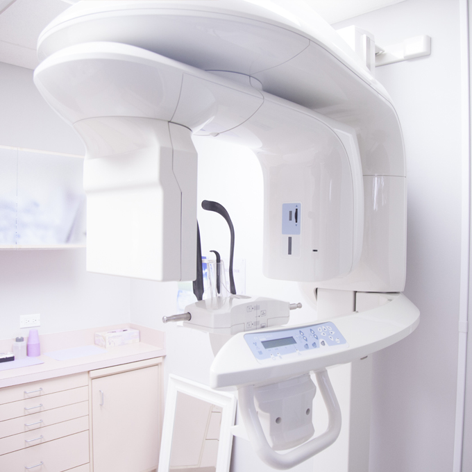
An example of a cbct scanner is i cat.
The x ray images are reconstructed by use of algorithms to come up with 3d high resolution images.
In office sinus ct scan cone beam ct scanner technology fairfax ent is proud to announce the availability of in office ct scanning for patients at its office.
This allows us to minimize radiation while providing a high tech three dimensional image of the sinuses.
Cone beam computed tomography is a medical imaging technique consisting of x ray computed tomography where the x rays are divergent forming a cone.
We are the only ent office in northern virginia with this new technology that provides a low radiation dose and in office convenience.
A cbct scanner uses a cone beam radiating from an x ray source in the shape of a cone covering large volume with one single rotation about the patient.
Perhaps because of the increased access to such technology cbct scanners are now finding many uses in dentistry such as in the fields of oral surgery endodontics and orthodontics.